Mini – PET Scanner Project
Introduction: What is a PET Scanner?
The Positron Emission Tomography (PET) is a non-invasive radioactive imaging technique aimed for the 3D reconstruction of a distributed positron sources. For being used in nuclear medicine, the tissues being probed should assimilate a radioisotope, this is a drug containing a sort half-life beta-plus decaying atom replacing a normal atom on its molecular structure.
The obtained images become valuable for study of metabolic processes and organs activity without the need of surgical intervention. Small tumors in an early cancer stage and brain disorders can be revealed, making the healing treatment more effective.

Figure: In nuclear medicine imaging, radio-pharmaceuticals with affinity to the tissue to be analyzed are taken by the patient. The array of detectors of the PET scanner detect the radiation emmited and creates data with which a 3d image of the tissue is generated.
Working principle
After the emmision, the positron travels a few milimiter suffering elastic scattering and loosing kinetic energy before anihilate with an electron present in the tissue. The rest energy of those particles is used to create two gamma photons, propagating in opposite direction and carring 511 keV energy each one.
The PET scanner, composed by and array of scintillators, and photon counters should be able to detect the two emmited photons and the arrival time of each one, in order to identify the anihilation point in the line of response. Data collected is used to reconstruct, with a few milimiters precision the shape of the tissue which absorbed the radiopharmaceutical.
Beta plus decaying element, such as carbon-11, fluorine-18, nitrogen-13, could be used in the artificial synthesis of organic molecules like suggars, aminoacids or neurotransmissors, so biological processes inside organs can be probed, after the radiopharmaceutical is assimilated.
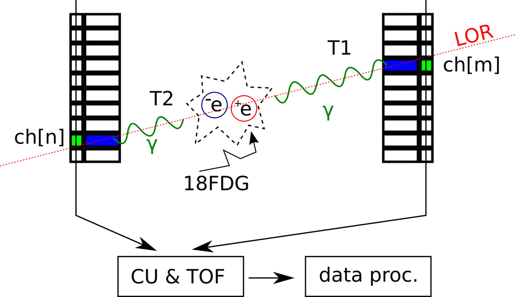
Figure: The array of detectors of the PET Scanner must be capable of detecting simultaneously the energy deposited by the pair of photons emitted after the decay of a radiopharmaceuticals that emits positrons. Also, it should determine the line of response (LOR) and the difference between arrival times of the photons. The collected information is processed to find out where did the electron/positron annihilation occurred and describe the distribution of the radioactive source.
Work Plan
In a first stage the goal is to construct a mini PET scanner, using same instrumentation as in high energy physics, like scintillating radiation detectors, WLS fibers, solid state photon counters and pulse processing electronics. Milestones are
- Construction of hybrid light guides containing wavelength shiffter (WLS) core and extra cladding.
- Micrometric description of two LYSO crystal matrices using dynascope imaging, for obtaining relevant geometrical parameters for design of light collector layers.
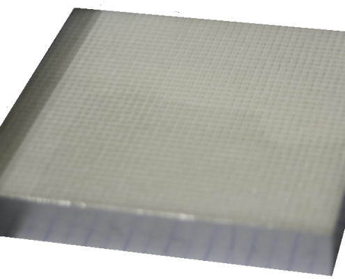
Figure: Isometric view of the visual characterization of the scintillating cristal matrices.
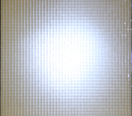
Figure: Front view of the visual characterization of the scintillating cristal matrices.
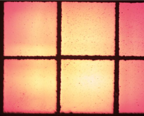
Figure: Microscopic view of some of the crystals inside the matrix.
3. Experimental research on light yield of different preparation of WLS, exciting with blue light source emulating the LYSO luminiscence. PIN diode and MPPC responses are going to be used to determine the best yi
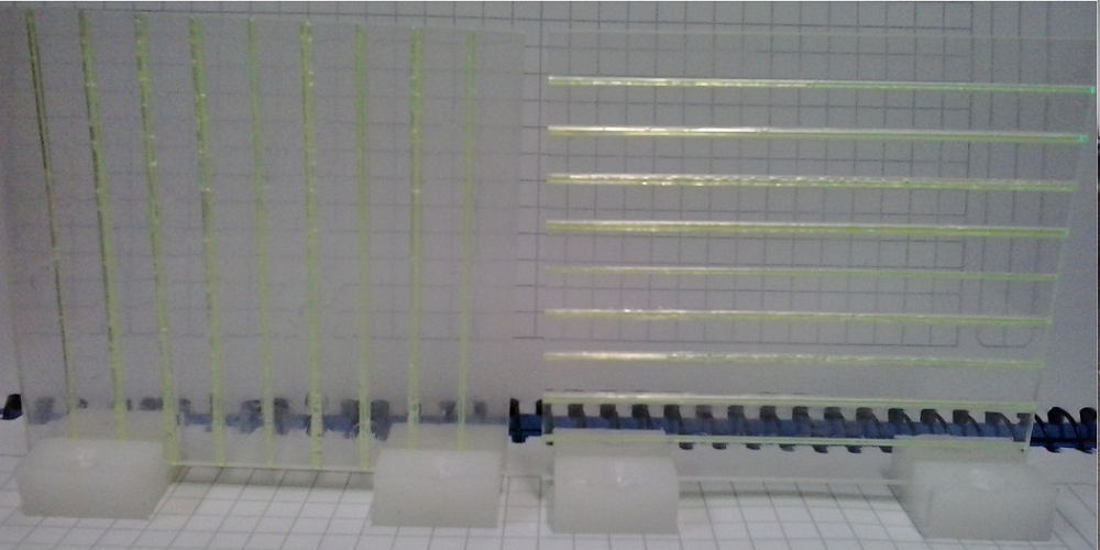
Figure: Light collectors fabricated in the laboratory.

Figure: Individual illumination of light collectors.
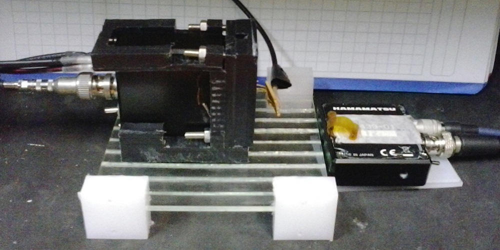
Figure: Light yield measured using PIN diodes.
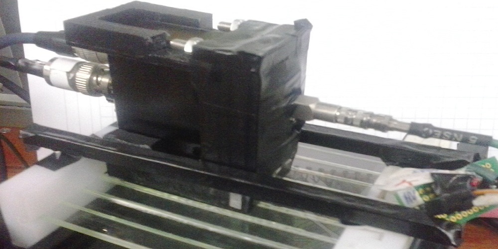
Figure: Light yield measured using MPPC and PIN diode.
4. Detect the light emmited by the LYSO matrices exposed to cosmic muons and gamma rays from Na22 decaying. Determine their response and energy uncertainty.
5. Optimization of geometrical parameters for improve light yield.
6. Write a Technical Design Report.
7. Build the final light collector grid, fix MPPC’s, connect to preamplifier electronics and to discriminator/TOF units for collecting data using a know radiation source.
